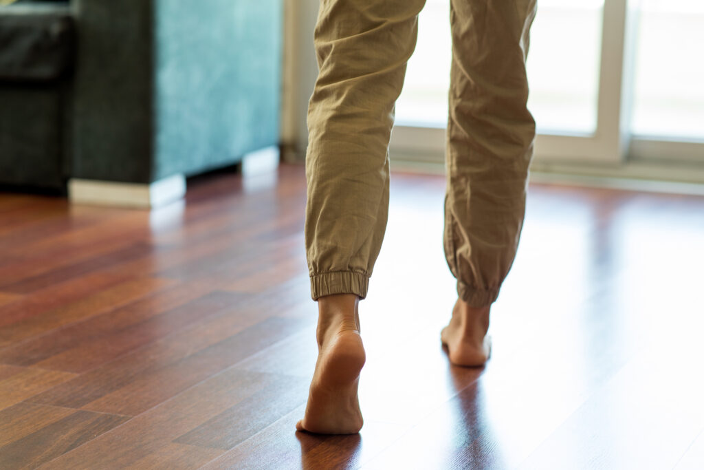
Your Trusted 24 Hour Orthopedic Care Provider

Services

Foot and Ankle

Containing more than 100 bones, tendons, muscles and ligaments and forming 33 joints, our feet and ankles play a major role in how we can (or can’t) move. A mild sprain or strain or even a broken toe can cause pain, limit our movement or possibly cause further injury in other parts of the body.

From bunions and heel problems to fractures and sprains, Orthopedic Associates’ fellowship-trained surgeons offer surgical techniques and non-surgical treatments for adults and children to eliminate or relieve many of these problems.

Trust our team of foot and ankle specialists to help you get back on your feet!


Tips for Healthy Feet:

Meet our

Foot and Ankle Team
William Barrick, M.D.
ORTHOPEDIC SPINE SURGEON / TRAUMA / GENERAL ORTHOPEDICS
Richard Dentico, M.D.
PAIN MANAGEMENT / REGENERATIVE MEDICINE / VEIN DISEASE
Daniel Kelmanovich M.D.
ORTHOPEDIC FOOT & ANKLE SURGEON
Lawrence Kusior, M.D.
ORTHOPEDIC SURGEON / SPORTS MEDICINE
Kenneth K. Rauschenbach, D.O.
KNEE, HIP & SHOULDER REPLACEMENT SURGEON / SPORTS MEDICINE / GENERAL ORTHOPEDICS / TRAUMA
Brian Reade, DPM
FOOT & ANKLE SURGEON / PODIATRIST
Vishal P. Rekhala, D.O.
SPECIALIZING IN PAIN MANAGEMENT / VEIN DISEASE / REGENERATIVE MEDICINE
Wen Shen, M.D.
ORTHOPEDIC FOOT & ANKLE SURGEON
Karl Ziermann, D.O.
SPORTS MEDICINE / CONCUSSION MANAGEMENT SPECIALIST / OSTEOPOROSIS MANAGEMENT SPECIALIST Schedule
Jean Walsh, MS, FNP-C, CEN
Landon Brown, MD
ORTHOPEDIC KNEE & HIP REPLACEMENT SURGEON

Conditions

Platelet Rich Plasma (PRP) is a non-operative solution in treating musculoskeletal, sports, and orthopedics injuries. PRP is concentrated from your own blood to trigger healing/repair process in the area of injury.

In PRP, the platelet is used as the initiator of repair. Platelets can release a multitude of signaling proteins, growth factors, chemotactic factors which stimulate tissue repair and vascular remodeling. This cascade of events allows the tissue to return to normal structure improving function and pain.

What are the benefits of PRP Treatment?
PRP therapy shows great promise in restoration of normal soft tissue architecture and regeneration of tissue such as tendons and ligaments. Patients can expect to see significant improvement in symptoms and eliminate the need for other treatments such as cortisone injections, medications or surgery.

Little to no Risk Factors
PRP is from your own blood, so there little to no risk factors with this treatment. Side effects or complications with PRP are very rare.

PRP may be effectively used in the treatment of:
  • Sports Injuries
  • Arthritic Joints
  • Lower Back Pain
  • Degenerative Disc Disease
  • Tennis Elbow
  • Golfer’s Elbow
  • Carpal Tunnel Syndrome
  • ACL Injuries
  • Shin Splints
  • Rotator Cuff Injuries
  • Plantar Fasciitis
  • Iliotibial Band (ITB) Syndrome
  • Achilles Tendinopathy
Prolotherapy treatment commonly consists of several injection sessions delivered every two to six weeks over the course of several months. During an individual prolotherapy session, therapeutic solutions are injected at sites of painful and tender ligament and tendon insertions, and in adjacent joint spaces. Injected solutions (“proliferants”) have historically been hypothesized to cause local irritation with subsequent inflammation and tissue healing, resulting in enlargement and strengthening of damaged ligamentous, tendon and intra-articular structures. These processes were thought to improve joint stability, biomechanics, function and ultimately, to decrease pain.

Prolotherapy may be effectively used in the treatment of:
  • Painful Degenerative Tendon Conditions
  • (i.e. Tendinopathies of the Hips, Rotator Cuff Tendons & Elbow)
  • Osteoarthritis
  • Plantar Fasciitis
  • Elbow Pain
  • Medial/Lateral Epicondylitis
  • Ulnar Ligament Strains
  • Chronic Neck and Low Back Pain
  • Sacroiliac Pain
  • Chronic Lateral Hip Pain
  • Coccydynia or Tailbone Pain
  • Osteitis Pubis
  • Ankle Pain and Instability
  • TMJ Dysfunction

Regenerative Cellular Therapies, or Concentrated Bone Marrow Aspirate, contains all of the healing and growth factors contained in PRP, with additional healing cells called pluripotent cells.

Bone marrow is the tissue that is found in the hollow spaces in the interior of our bones. It is known that significant sources of our body’s adult stem cells are contained within our bone marrow. It is also known that these have the potential of positively contributing to healing. Blood and tissue can be harvested from the several bones in the body, the most popular being the hip, tibia and calcaneus, in an outpatient setting via an aspiration device.

Similar to PRP, a centrifugation device is used to concentrate the platelets and growth factors, as well as the pluripotent (or stem) cells, creating an injectable product that is delivered directly to the site of injury, jump-starting the healing cascade.

Regenerative Cellular Therapies may be effectively used in the treatment of:

  • Muscle Injuries
  • Bicipital Tendonitis
  • Ankle Sprain/Ligament Pain
  • Painful Degenerative Tendon Conditions
  • (i.e. Tendinopathies of the Hips, Rotator Cuff Tendons & Elbow)
  • Osteoarthritis
  • Plantar Fasciitis
  • Elbow Pain
  • Medial/Lateral Epicondylitis
  • Ulnar Ligament Strains
  • Chronic Neck and Low Back Pain
  • Sacroiliac Pain
  • Chronic Lateral Hip Pain
  • Coccydynia or Tailbone Pain
  • Osteitis Pubis
  • Ankle Pain and Instability
  • TMJ Dysfunction
Arthritis is inflammation of one or more of your joints. It can cause pain and stiffness in any joint in the body, and is common in the small joints of the foot and ankle.

There are more than 100 forms of arthritis, many of which affect the foot and ankle. All types can make it difficult to walk and perform activities you enjoy.

Although there is no cure for arthritis, there are many treatment options available to slow the progress of the disease and relieve symptoms. With proper treatment, many people with arthritis are able to manage their pain, remain active, and lead fulfilling lives.

Symptoms

The symptoms of arthritis vary depending on which joint is affected. In many cases, an arthritic joint will be painful and inflamed. Generally, the pain develops gradually over time, although sudden onset is also possible. There can be other symptoms, as well, including:
  • Pain with motion
  • Pain that flares up with vigorous activity
  • Tenderness when pressure is applied to the joint
  • Joint swelling, warmth, and redness
  • Increased pain and swelling in the morning, or after sitting or resting
  • Difficulty in walking due to any of the above symptoms

A bunion is a painful bony bump that develops on the inside of the foot at the big toe joint. Bunions are often referred to as hallux valgus.

Bunions develop slowly. Pressure on the big toe joint causes the big toe to lean toward the second toe. Over time, the normal structure of the bone changes, resulting in the bunion bump. This deformity will gradually increase and may make it painful to wear shoes or walk.

Anyone can get a bunion, but they are more common in women. Many women wear tight, narrow shoes that squeeze the toes together-which makes it more likely for a bunion to develop, worsen and cause painful symptoms.

In most cases, bunion pain is relieved by wearing wider shoes with adequate toe room and using other simple treatments to reduce pressure on the big toe.

Symptoms

In addition to the visible bump on the inside of the foot, symptoms of a bunion may include:

  • Pain and tenderness
  • Redness and inflammation
  • Hardened skin on the bottom of the foot
  • A callus or corn on the bump
  • Stiffness and restricted motion in the big toe, which may lead to difficulty in walking

A fracture of the calcaneus, or heel bone, can be a painful and disabling injury. This type of fracture commonly occurs during a high-energy event-such as a car crash or a fall from a ladder-when the heel is crushed under the weight of the body. When this occurs, the heel can widen, shorten, and become deformed.

Calcaneus fractures can be quite severe. Treatment often involves surgery to reconstruct the normal anatomy of the heel and restore mobility so that patients can return to normal activity. But even with appropriate treatment, some fractures may result in long-term complications, such as pain, swelling, loss of motion, and arthritis.

Symptoms

Patients with calcaneus fractures usually experience:

  • Pain
  • Bruising
  • Swelling
  • Heel deformity
  • Inability to put weight on the heel or walk


With some minor calcaneus fractures, the pain may not be enough to prevent you from walking – but you may limp. This is because your Achilles tendon acts through the calcaneus to support your body weight. If, however, your calcaneus is deformed by the injury, your muscle and tendon cannot generate enough power to support your weight. Your foot and ankle will feel unstable, and you will walk differently.

Charcot-Marie-Tooth disease is a group of disorders that affect the peripheral nerves – the nerves that carry messages between the brain and muscles throughout the body. It is named after the three doctors who described it in 1886: Jean Martin Charcot and Pierre Marie in Paris, and Howard Henry Tooth in Cambridge, England. Charcot-Marie-Tooth disease is also sometimes referred to as hereditary motor and sensory neuropathy (HMSN) or peroneal muscular atrophy.

All types of Charcot-Marie-Tooth disease (CMT) cause degeneration of the peripheral nerves, leading to muscle weakness and some loss of sensation in the arms, legs, hands, and feet. These symptoms often first appear during adolescence or early adulthood, but can develop later in life, as well.

Symptoms vary greatly among people with CMT, and usually begin in the feet and legs. As CMT progresses, it can cause deformities in the bones of the feet, such as hammertoes and high arches. Without treatment, walking may become difficult.

Although there is no cure for Charcot-Marie-Tooth disease, there are many treatment options and assistive devices to help people manage physical challenges and lead fulfilling lives.

Symptoms

People with CMT have varied symptoms, depending upon the type of CMT they have, as well as the severity of their disease. In general, symptoms fall into three major categories:
  • Muscle weakness
  • Bone deformity
  • Loss of sensation

Most people first notice CMT symptoms in their feet and legs. This is a slowly progressive disorder. Over time, symptoms may also affect the hands and arms.
People often blame the common foot deformity claw toe on wearing shoes that squeeze your toes, such as shoes that are too short or high heels. However, claw toe also is often the result of nerve damage caused by diseases like diabetes or alcoholism, which can weaken the muscles in your foot. Having claw toe means your toes “claw,” digging down into the soles of your shoes and creating painful calluses. Claw toe gets worse without treatment and may become a permanent deformity over time.

Symptoms
  • Your toes are bent upward (extension) from the joints at the ball of the foot.
  • Your toes are bent downward (flexion) at the middle joints toward the sole of your shoe.
  • Sometimes your toes also bend downward at the top joints, curling under the foot.
  • Corns may develop over the top of the toe or under the ball of the foot.
Compartment syndrome is a painful condition that occurs when pressure within the muscles builds to dangerous levels. This pressure can decrease blood flow, which prevents nourishment and oxygen from reaching nerve and muscle cells.

Compartment syndrome can be either acute or chronic.

Acute compartment syndrome is a medical emergency. It is usually caused by a severe injury. Without treatment, it can lead to permanent muscle damage.

Chronic compartment syndrome, also known as exertional compartment syndrome, is usually not a medical emergency. It is most often caused by athletic exertion.

Symptoms

Acute Compartment Syndrome
The classic sign of acute compartment syndrome is pain, especially when the muscle within the compartment is stretched.
  • The pain is more intense than what would be expected from the injury itself. Using or stretching the involved muscles increases the pain.
  • There may also be tingling or burning sensations (paresthesias) in the skin.
  • The muscle may feel tight or full.
  • Numbness or paralysis are late signs of compartment syndrome. They usually indicate permanent tissue injury.

Chronic (Exertional) Compartment Syndrome
Chronic compartment syndrome causes pain or cramping during exercise. This pain subsides when activity stops. It most often occurs in the leg.
Symptoms may also include:

  • Numbness
  • Difficulty moving the foot
  • Visible muscle bulging
Every day, the average person spends several hours on their feet and takes several thousand steps. Walking puts pressure on your feet that’s equivalent to 2-3 times your body weight. No wonder your feet hurt!

Actually, most foot problems can be blamed not on walking but on your walking shoes. Corns, for example, are calluses that form on the toes because the bones push up against the shoe and put pressure on the skin. The surface layer of the skin thickens and builds up, irritating the tissues underneath. Hard corns are usually located on the top of the toe or on the side of the small toe. Soft corns resemble open sores and develop between the toes as they rub against each other.

Cause

  • Shoes that don’t fit properly. If shoes are too tight, they squeeze the foot, increasing pressure. If they are too loose, the foot may slide and rub against the shoe, creating friction.
  • Toe deformities, such as hammer toe or claw toe.
  • High heeled shoes because they increase the pressure on the forefoot.
  • Rubbing against a seam or stitch inside the shoe.
  • Socks that don’t fit properly.
Diabetes is a condition of elevated blood sugar that affects about 6 percent of the population in the United States, or about 16 million people. Diabetic foot problems are a major health concern and are a common cause of hospitalization.

Most foot problems that people with diabetes face arise from two serious complications of the disease: nerve damage and poor circulation. One of the more critical foot problems these complications can cause is Charcot arthropathy, which can deform the shape of the foot and lead to disability.

There are treatment options for the wide range of diabetic foot problems. The most effective treatment, however, is prevention. For people with diabetes, careful, daily inspection of the feet is essential to overall health and the prevention of damaging foot problems.

Symptoms

Although a patient with Charcot arthropathy typically will not have much pain, they may have other symptoms.
  • The most sensitive sign of early Charcot foot is swelling of the foot. This can occur without an obvious injury.
  • Redness of the foot can also occur in the early stages.
  • The swelling, redness, and changes to the bone that are seen on x-ray may be confused for a bone infection. A bone infection is very unlikely if the skin is intact and there is no ulcer present.

A hammer toe is a deformity of the second, third or fourth toes. In this condition, the toe is bent at the middle joint, so that it resembles a hammer. Initially, hammer toes are flexible and can be corrected with simple measures but, if left untreated, they can become fixed and require surgery.

People with hammer toe may have corns or calluses on the top of the middle joint of the toe or on the tip of the toe. They may also feel pain in their toes or feet and have difficulty finding comfortable shoes.

Cause

Hammer toe results from shoes that don’t fit properly or a muscle imbalance, usually in combination with one or more other factors. Muscles work in pairs to straighten and bend the toes. If the toe is bent and held in one position long enough, the muscles tighten and cannot stretch out.

Shoes that narrow toward the toe may make your forefoot look smaller. But they also push the smaller toes into a flexed (bent) position. The toes rub against the shoe, leading to the formation of corns and calluses, which further aggravate the condition. A higher heel forces the foot down and squishes the toes against the shoe, increasing the pressure and the bend in the toe. Eventually, the toe muscles become unable to straighten the toe, even when there is no confining shoe.

Every mile you walk puts 60 tons of stress on each foot. Your feet can handle a heavy load, but too much stress pushes them over their limits. When you pound your feet on hard surfaces playing sports or wear shoes that irritate sensitive tissues, you may develop heel pain, the most common problem affecting the foot and ankle.

A sore heel will usually get better on its own without surgery if you give it enough rest. However, many people try to ignore the early signs of heel pain and keep on doing the activities that caused it. When you continue to use a sore heel, it will only get worse and could become a chronic condition leading to more problems. Surgery is rarely necessary.

If you trim your toenails too short, particularly on the sides of your big toes, you may set the stage for an ingrown toenail. Like many people, when you trim your toenails, you may taper the corners so that the nail curves with the shape of your toe. But this technique may encourage your toenail to grow into the skin of your toe. The sides of the nail curl down and dig into your skin. An ingrown toenail may also happen if you wear shoes that are too tight or too short.

Symptoms

When you first have an ingrown toenail, it may be hard, swollen and tender. Later, it may get red and infected, and feel very sore. Ingrown toenails are a common, painful condition-particularly among teenagers. Any of your toenails can become ingrown, but the problem more often affects the big toe. An ingrown nail occurs when the skin on one or both sides of a nail grows over the edges of the nail, or when the nail itself grows into the skin. Redness, pain and swelling at the corner of the nail may result and infection may soon follow. Sometimes a small amount of pus can be seen draining from the area.

Ingrown nails may develop for many reasons. Some cases are congenital-the nail is just too large for the toe. Trauma, such as stubbing the toe or having the toe stepped on, may also cause an ingrown nail. However, the most common cause is tight shoe wear or improper grooming and trimming of the nail.

Lisfranc (midfoot) injuries result if bones in the midfoot are broken or ligaments that support the midfoot are torn. The severity of the injury can vary from simple to complex, involving many joints and bones in the midfoot.

A Lisfranc injury is often mistaken for a simple sprain, especially if the injury is a result of a straightforward twist and fall. However, injury to the Lisfranc joint is not a simple sprain that should be simply “walked off.” It is a severe injury that may take many months to heal and may require surgery to treat.

Symptoms

The most common symptoms of Lisfranc injury include:
  • The top of foot may be swollen and painful.
  • There may be bruising on both the top and bottom of the foot. Bruising on the bottom of the foot is highly suggestive of a Lisfranc injury.
  • Pain that worsens with standing, walking or attempting to push off on the affected foot. The pain can be so severe that crutches may be required.

If standard treatment for a sprain (rest, ice, elevation) does not relieve pain and swelling, you should seek care from an orthopaedic surgeon.

Conditions

If you sometimes feel that you are “walking on a marble,” and you have persistent pain in the ball of your foot, you may have a condition called Morton’s neuroma. A neuroma is a benign tumor of a nerve. Morton’s neuroma is not actually a tumor, but a thickening of the tissue that surrounds the digital nerve leading to the toes.

Symptoms
  • Normally, there are no outward signs, such as a lump, because this is not really a tumor.
  • Burning pain in the ball of the foot that may radiate into the toes. The pain generally intensifies with activity or wearing shoes. Night pain is rare.
  • There may also be numbness in the toes, or an unpleasant feeling in the toes.

Runners may feel pain as they push off from the starting block. High-heeled shoes, which put the foot in a similar position to the push-off, can also aggravate the condition. Tight, narrow shoes also aggravate this condition by compressing the toe bones and pinching the nerve.
This article addresses pilon fractures-a specific type of fracture that occurs in the lower leg near the ankle. To find in-depth information on ankle fractures, please read Ankle Fractures (Broken Ankle).

A pilon fracture is a type of break that occurs at the bottom of the tibia (shinbone) and involves the weight-bearing surface of the ankle joint. With this type of injury, the other bone in the lower leg, the fibula, is frequently broken as well. A pilon fracture typically occurs as the result of a high-energy event, such as a car collision or fall from height.

Pilon is the French word for “pestle”-an instrument used for crushing or pounding. In many pilon fractures, the bone may be crushed or split into several pieces due to the high-energy impact that caused the injury.

In most cases, surgery is needed to restore the damaged bone to its normal position. Because of the energy required to cause a pilon fracture, patients may have other injuries that require treatment as well.

Symptoms

Patients with pilon fractures usually experience immediate and severe pain. Other symptoms may include:
  • Swelling
  • Bruising
  • Tenderness
  • Inability to bear weight on the injured leg
  • Deformity-your ankle may look angled or crooked
Plantar fasciitis (fashee-EYE-tiss) is the most common cause of pain on the bottom of the heel. Approximately 2 million patients are treated for this condition every year.

Plantar fasciitis occurs when the strong band of tissue that supports the arch of your foot becomes irritated and inflamed.

Symptoms

The most common symptoms of plantar fasciitis include:
  • Pain on the bottom of the foot near the heel
  • Pain with the first few steps after getting out of bed in the morning, or after a long period of rest, such as after a long car ride. The pain subsides after a few minutes of walking
  • Greater pain after (not during) exercise or activity
Posterior tibial tendon dysfunction is one of the most common problems of the foot and ankle. It occurs when the posterior tibial tendon becomes inflamed or torn. As a result, the tendon may not be able to provide stability and support for the arch of the foot, resulting in flatfoot.

Most patients can be treated without surgery, using orthotics and braces. If orthotics and braces do not provide relief, surgery can be an effective way to help with the pain. Surgery might be as simple as removing the inflamed tissue or repairing a simple tear. However, more often than not, surgery is very involved, and many patients will notice some limitation in activity after surgery.

Symptoms
  • Pain along the inside of the foot and ankle, where the tendon lies. This may or may not be associated with swelling in the area.
  • Pain that is worse with activity. High-intensity or high-impact activities, such as running, can be very difficult. Some patients can have trouble walking or standing for a long time.
  • Pain on the outside of the ankle. When the foot collapses, the heel bone may shift to a new position outwards. This can put pressure on the outside ankle bone. The same type of pain is found in arthritis in the back of the foot.
Rheumatoid arthritis is a chronic disease that attacks multiple joints throughout the body. It most often starts in the small joints of the hands and feet, and usually affects the same joints on both sides of the body.

More than 90% of people with rheumatoid arthritis (RA) develop symptoms in the foot and ankle over the course of the disease.

Symptoms

The most common symptoms are pain, swelling, and stiffness. Unlike osteoarthritis, which typically affects one specific joint, symptoms of RA usually appear in both feet, affecting the same joints on each foot.

Most bones in the human body are connected to each other at joints. But there are a few bones that are not connected to any other bone. Instead, they are connected only to tendons or are embedded in muscle. These are the sesamoids. The kneecap (patella) is the largest sesamoid. Two other very small sesamoids (about the size of a kernel of corn) are found in the underside of the forefoot near the great toe, one on the outer side of the foot and the other closer to the middle of the foot.

Sesamoids act like pulleys. They provide a smooth surface over which the tendons slide, thus increasing the ability of the tendons to transmit muscle forces. The sesamoids in the forefoot also assist with weightbearing and help elevate the bones of the great toe. Like other bones, sesamoids can break (fracture). Additionally, the tendons surrounding the sesamoids can become irritated or inflamed. This is called sesamoiditis and is a form of tendinitis. It is common among ballet dancers, runners and baseball catchers.

Symptoms
  • Pain is focused under the great toe on the ball of the foot. With sesamoiditis, pain may develop gradually; with a fracture, pain will be immediate.
  • Swelling and bruising may or may not be present.
  • You may experience difficulty and pain in bending and straightening the great toe.
Sever’s disease (also known as calcaneal apophysitis) is one of the most common causes of heel pain in growing children and adolescents. It is an inflammation of the growth plate in the calcaneus (heel).

Sever’s disease is caused by repetitive stress to the heel, and most often occurs during growth spurts, when bones, muscles, tendons, and other structures are changing rapidly. Children and adolescents who participate in athletics-especially running and jumping sports-are at an increased risk for this condition. However, less active adolescents may also experience this problem, especially if they wear very flat shoes.

In most cases of Sever’s disease, simple measures like rest, over-the-counter medication, a change in footwear, and stretching and strengthening exercises will relieve pain and allow a return to daily activities.

Symptoms

Painful symptoms are often brought on by running, jumping, and other sports-related activities. In some cases, both heels have symptoms, although one heel may be worse than the other. Symptoms may include:
  • Heel pain and tenderness underneath the heel
  • Mild swelling at the heel
An ankle sprain occurs when the strong ligaments that support the ankle stretch beyond their limits and tear. Ankle sprains are common injuries that occur among people of all ages. They range from mild to severe, depending upon how much damage there is to the ligaments.

Most sprains are minor injuries that heal with home treatments like rest and applying ice. However, if your ankle is very swollen and painful to walk on – or if you are having trouble putting weight on your ankle at all, be sure to see your doctor.

Without proper treatment and rehabilitation, a more severe sprain can weaken your ankle-making it more likely that you will injure it again. Repeated ankle sprains can lead to long-term problems, including chronic ankle pain, arthritis, and ongoing instability.

Symptoms

A sprained ankle is painful. Other symptoms may include:
  • Swelling
  • Bruising
  • Tenderness to touch
  • Instability of the ankle-this may occur when there has been complete tearing of the ligament or a complete dislocation of the ankle joint.
The most common site of arthritis in the foot is at the base of the big toe. This joint is called the metatarsophalangeal, or MTP joint. It’s important because it has to bend every time you take a step. If the joint starts to stiffen, walking can become painful and difficult.

In the MTP joint, as in any joint, the ends of the bones are covered by a smooth articular cartilage. If wear-and-tear or injury damage the articular cartilage, the raw bone ends can rub together. A bone spur, or overgrowth, may develop on the top of the bone. This overgrowth can prevent the toe from bending as much as it needs to when you walk. The result is a stiff big toe, or hallux rigidus.

Hallux rigidus usually develops in adults between the ages of 30 and 60 years. No one knows why it appears in some people and not others. It may result from an injury to the toe that damages the articular cartilage or from differences in foot anatomy that increase stress on the joint.

Symptoms
  • Pain in the joint when you are active, especially as you push-off on the toes when you walk
  • Swelling around the joint
  • A bump, like a bunion or callus, that develops on the top of the foot
  • Stiffness in the great toe and an inability to bend it up or down

A stress fracture is a small crack in a bone, or severe bruising within a bone. Most stress fractures are caused by overuse and repetitive activity, and are common in runners and athletes who participate in running sports, such as soccer and basketball.

Stress fractures usually occur when people change their activities – such as by trying a new exercise, suddenly increasing the intensity of their workouts, or changing the workout surface (jogging on a treadmill vs. jogging outdoors). In addition, if osteoporosis or other disease has weakened the bones, just doing everyday activities may result in a stress fracture.

The weight-bearing bones of the foot and lower leg are especially vulnerable to stress fractures because of the repetitive forces they must absorb during activities like walking, running, and jumping.

Refraining from high impact activities for an adequate period of time is key to recovering from a stress fracture in the foot or ankle. Returning to activity too quickly can not only delay the healing process but also increase the risk for a complete fracture. Should a complete fracture occur, it will take far longer to recover and return to activities.

Symptoms

The most common symptom of a stress fracture in the foot or ankle is pain. The pain usually develops gradually and worsens during weight-bearing activity. Other symptoms may include:

  • Pain that diminishes during rest
  • Pain that occurs and intensifies during normal, daily activities
  • Swelling on the top of the foot or on the outside of the ankle
  • Tenderness to touch at the site of the fracture
  • Possible bruising

A talus fracture is a break in one of the bones that forms the ankle. This type of fracture often occurs during a high-energy event, such as a car collision or a high-velocity fall.

Because the talus is important for ankle movement, a fracture often results in significant loss of motion and function. In addition, a talus fracture that does not heal properly can lead to serious complications, including chronic pain. For this reason, many talus fractures require surgery.

Symptoms

Patients with talus fractures usually experience:

  • Acute pain
  • Inability to walk or bear weight on the foot
  • Considerable swelling, bruising, and tenderness

A tarsal coalition is an abnormal connection of two or more bones in the foot. The bones affected – called tarsal bones – are located toward the back of the foot and in the heel, and the connection of the bones can result in a severe, rigid flatfoot.

Although tarsal coalition is often present at birth, children typically do not show signs of the disorder until early adolescence. The foot may become stiff and painful, and everyday physical activities are often difficult.

For many children with tarsal coalition, symptoms are relieved with simple treatments, such as orthotics and physical therapy. If a child has severe symptoms that do not respond to simple treatments and continue to interfere with their daily activities, surgery may be recommended.

Symptoms

Many tarsal coalitions are never discovered because they do not cause symptoms or any obvious foot deformity. When symptoms do occur, they may include:

  • Stiff, painful feet. The pain usually occurs below the ankle around the middle or back half of the foot.
  • A rigid, flat foot that makes it difficult to walk on uneven surfaces. To accommodate for the foot’s lack of motion, the patient may roll the ankle more than normal, which may result in recurrent ankle sprains.
  • Increased pain or a limp with higher levels of activity.
Fractures of the toes and forefoot are quite common. Fractures can result from a direct blow to the foot-such as accidentally kicking something hard or dropping a heavy object on your toes. They can also result from the overuse and repetitive stress that comes with participating in high-impact sports like running and basketball.

Although fracturing a bone in your toe or forefoot can be quite painful-it rarely requires surgery. In most cases, a fracture will heal with rest and a change in activities.

Symptoms

The most common symptoms of a fracture are pain and swelling. Other symptoms may include:
  • Bruising or discoloration that extends to nearby parts of the foot
  • Pain with walking and weight bearing


Learn About Our Advanced Technologies

We offer advanced treatment options for patients with special needs.

Before beginning any treatment, discuss your options with your foot doctor.

Advanced Technologies

MLS Laser Therapy is an FDA-cleared treatment for pain that uses concentrated light energy to stimulate the body’s own healing process. Laser therapy minimizes pain and inflammation as an alternative to prescriptions and surgery. It reduces recovery times, which is especially important post-surgery, so patients can quickly return to a higher quality of life. MLS (multi-wave locked system) technology in specific delivers two therapeutic wavelengths – the 808nm (anti-edemic and anti-inflammatory) and the 905nm (analgesic). The combination of these wavelengths produces greater pain-relieving effects than either can produce on their own, while also minimizing the risk of thermal damage. It is this unique combination and synchronization of continuous and pulsed emissions that characterizes MLS and distinguishes it from other Class IV lasers.

Platelet-rich plasma (PRP) is a patient’s own concentrated platelets. PRP contains a large number growth factors. These growth factors stimulate healing. When PRP is injected, it can aid the body’s natural healing process. The goal is not only to relieve symptoms, but to also create actual healing. In some cases, PRP may reduce the need for medication and/or surgery. PRP has been used to treat tendon, ligament, cartilage and bone injuries, as well as arthritis. Around the foot and ankle, PRP is used for treatment of tendon and ligament injuries, such as plantar fasciitis, Achilles tendon and ankle ligament injuries.

A small amount of a patient’s blood is drawn and then spun at high speed. The platelets are concentrated. This liquid is then injected around or near the area of injury being treated. The PRP at this stage contains three to five times the concentration of growth factors compared to normal human blood.

What Happens After a PRP Injection?

You may experience mild pain and irritation of the area for several days following the injection. Some doctors may ask patients to limit motion or weight-bearing activity immediately following the injection. The use of a brace, boot or cast may be recommended during the early post-injection course.

Three to seven days after the injection, you may gradually return to normal physical activities. The return to full activity is determined based on response to the therapy and the recommendation of your surgeon.

The entire process takes only 20 minutes and is performed in a hospital using intravenous sedation anesthetic (you are asleep but breathing on your own). Your doctor places the Topaz wand, which looks like a sewing needle, into the affected fascia or tendon for just a half-second. Then, the wand is moved about a quarter-inch and pushed in again. This action is repeated until a grid-like pattern is formed around the affected area.

The Topaz wand uses cobalation technologies, which are radio frequency waves that gently cause microscopic “trauma” to the scar tissue your body hasn’t yet been able to heal. The procedure brings blood back to the affected area and restarts the healing process by increasing the growth factors in the damaged tissue region. The strategically placed Topaz wand greatly minimizes any damage to surrounding healthy tissue.
Our Bracing Service supports surgical procedures, nonsurgical rehabilitation and pain management with its superior products and care. We offer this service at all of our office locations.
This procedure is for patients with chronic fascia and tendon problems, including tennis elbow, golfers’ elbow, patellar tendonitis, Achilles tendonitis and planter fasciitis.

Tenex procedure requires local anesthesia, an ultrasound and a tiny incision. A thin probe uses ultrasonic energy to remove the damaged portion of the tissue. The procedure takes 25 minutes.

Radiofrequency Nerve Ablation (RFA) is a safe, proven means of treating chronic heel pain. RFA is recommended for patients who have pain for over three months, and all other forms of therapy have been unresponsive.

How Radiofrequency Nerve Ablation Works

Radiofrequency energy is transmitted to the tip of a needle where it is converted to heat and targeted at specific nerve tissue. Once the nerves undergo this heat treatment, they slowly stop transmitting pain. Radiofrequency refers to radio waves, a form of electromagnetic energy produced during the procedure.

What to Expect After Your Radiofrequency Nerve Ablation Procedure

The RFA heel pain procedure takes 15 to 30 minutes and is performed on an outpatient basis. After the treatment is completed, a small bandage is placed over the probe insertion site, and you can return to normal activity almost immediately.

Chronic ankle instability is a condition characterized by a recurring “giving way” of the outer (lateral) side of the ankle. This condition often develops after repeated ankle sprains. Usually the “giving way” occurs while walking or doing other activities, but it can also happen while you’re just standing. Many athletes and ordinary people suffer from chronic ankle instability. People with chronic ankle instability often complain of: Repeated turning of the ankle, especially on uneven surfaces or when participating in sports Persistent (chronic) discomfort and swelling Pain or tenderness Chronic ankle instability usually develops following an ankle sprain that has not adequately healed or was not rehabilitated completely. When you sprain your ankle, the connective tissues (ligaments) are stretched or torn. The ability to balance is often affected. Proper rehabilitation is needed to strengthen the muscles around the ankle and “retrain” the tissues within the ankle that affect balance. If you suffer from chronic ankle instability ask your doctor about an internal brace for ligament repair.

Internal Brace for Ankle Ligament Repair

InternalBrace is a tiny, thick, rope-like fiber that’s stronger than the natural ligament. The fiber is secured in place by a BioComposite SwiveLock anchor, designed to allow blood and bone marrow to circulate through the device. The InternalBrace tightly joins the torn ligaments together to strengthen the repair and increase healing time. When their job is done, the fiber and anchors biodegrade and reabsorb into the body.
Although the foot and ankle community generally recognize the Brostrom procedure as the “gold standard” of ligament repair, augmenting the repair with an InternalBrace enhances stability and decreases recovery times. You’ll spend less time in a cast, start physical therapy sooner and return to sports much earlier than you would with a traditional repair.
With the minimally-invasive InternalBrace repair system, patients can expect to be out of their cast and into a walking boot in two to three weeks. You can start physical therapy as soon as you’re out of your cast. Although each case is different, patients generally spend less time in physical therapy thanks to the added stability offered by the InternalBrace. The sooner you’re out of physical therapy, the sooner you can get back on track toward your regular training routine.

Posted in on February, 2022